
Posted 12 February 2025

An international team of researchers has, for the first time, created a detailed map of the location and identity of individual cells in the adult lung, both from healthy lungs and in lungs from people affected by chronic lung disease.
A spatial map of gene expression in 1.6 million cells from the lungs of people with idiopathic pulmonary fibrosis (IPF) revealed a surprising discovery: some lung tissue in these patients shows signs of the disease before significant structural remodelling of the tissue occurs.
The AI-driven finding could point to future therapeutic strategies that treat IPF patients based on their individual stage of cellular and molecular remodelling.
The findings give hope that scientists may be able to halt or reverse disease by targeting early molecular changes before patients suffer substantial loss of lung function and develop debilitating symptoms from IPF.
People with IPF initially experience shortness of breath. IPF can progress very quickly and, without intervention, is fatal. Over 1,250 Australians are diagnosed with IPF every year, most of whom are between the ages of 50 and 70 years old.
“If you imagine a city map in an atlas, we’ve been able to create a ‘google street view’ to identify individual neighbourhoods and buildings, it’s that revolutionary,” said Associate Professor Davis McCarthy, one of the lead researchers on the study.
The study published in Nature Genetics used image-based spatial transcriptomics, an important tool for studying IPF and relies on artificial intelligence (AI) computational methods.
The team profiled 343 genes in lung tissue samples from 26 people who underwent lung transplant for PF and from nine people without PF.
“While there are some new treatments that can slow the progress of the disease, the only current effective therapy is lung transplantation, which has limited availability and in which one set of medical challenges are swapped for another,” said Associate Professor McCarthy, who co-led the research with his collaborator Associate Professor Nicholas Banovich from the Translational Genomics Research Institute and Associate Professor Jon Kropski at Vanderbilt University Medical Center in the U.S.
Their analysis helped them map out where cells with signs of PF occur, and identify the cellular and molecular underpinnings of some features of PF. They also characterized 12 distinct, molecularly defined spatial niches in healthy and PF-affected lungs.
“In recent years, it has become clear that genetic factors contribute substantially to a person’s risk of developing IPF, but the exact influence of these genetic variations has not been well understood,” said Associate Professor McCarthy.
-
Background Info
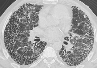
Each of the 300 million or so sacs in a healthy set of lungs are surrounded by capillaries that facilitate the transfer of the oxygen into the bloodstream. For this process to occur, the tissue that makes up the lungs must be elastic and flexible, able to be fully inflated with each breath. In someone with idiopathic pulmonary fibrosis (IPF), the thin lacy walls of the air sacs become stiffened and thick with scar tissue, and unable to be filled with air. Idiopathic means unexplained. We use the term when we can’t pinpoint a cause for a condition.
Image-based spatial transcriptomics is a cutting-edge technique that allows researchers to visualize and analyse gene expression in tissue samples while preserving spatial context. Unlike bulk RNA sequencing, which loses information about where in the tissue a gene is expressed, image-based spatial transcriptomics combines microscopy with molecular biology to map gene activity directly onto tissue sections. AI plays a crucial role in image-based spatial transcriptomics. The datasets generated by these techniques are vast and complex, requiring AI-driven computational methods to process, analyse, and interpret the spatial gene expression data.